Introduction
Juvenile Nasopharyngeal Angiofibroma (JNA) is a rare, benign but locally aggressive vascular tumor that primarily affects adolescent males between the ages of 10 and 25. Though histologically benign (non-cancerous), the tumor tends to grow aggressively, invading nearby structures such as the orbit, sinuses, skull base, and intracranial regions. Its vascular nature makes management particularly challenging due to the risk of heavy bleeding during surgery.
Pathogenesis and Risk Factors
The exact cause of JNA is not fully understood, but it is thought to originate from the sphenopalatine foramen, an area at the back of the nasal cavity. Hormonal factors, particularly the influence of androgens, are believed to play a role, explaining the tumor’s predominance in young males. There is no strong hereditary or environmental association, although some studies suggest a possible link with genetic mutations in growth-related pathways.
Symptoms
JNA grows slowly but can cause significant symptoms due to its size and location. The most common presenting features include:
- Nasal obstruction (often unilateral, progressing to bilateral)
- Recurrent, severe epistaxis (nosebleeds)
- Facial swelling or deformity if the tumor expands into adjacent areas
- Proptosis (bulging of the eye) if it invades the orbit
- Headaches or facial pain
- Hearing loss or ear fullness due to obstruction of the Eustachian tube
If untreated, the tumor may extend into the cranial cavity, leading to neurological symptoms such as vision problems, confusion, or even seizures.
Diagnosis
Diagnosing JNA requires a combination of clinical evaluation and imaging techniques:
- Clinical Examination:
- Nasal endoscopy can reveal a reddish, vascular mass in the posterior nasal cavity. However, biopsies are generally avoided due to the risk of excessive bleeding.
- Imaging Studies:
- CT Scan: Useful for evaluating the extent of bony involvement, particularly the erosion of the skull base or sinuses.
- MRI: Provides better visualization of soft tissue structures and tumor extension into the brain or orbit.
- Angiography: Often performed to assess the vascular supply, particularly from the external carotid artery, and helps guide pre-surgical embolization.
Staging of JNA
Several classification systems exist, but the Radkowski system is widely used for staging JNA based on the extent of tumor spread:
- Stage I: Limited to the nasal cavity and nasopharynx.
- Stage II: Extends into the pterygoid fossa or sinuses without skull base erosion.
- Stage III: Erodes the skull base or invades the orbit.
- Stage IV: Intracranial extension with or without brain invasion.
Staging helps guide treatment and predict outcomes.
Management
Treatment of JNA is complex due to the tumor’s vascularity, aggressive growth, and involvement of critical anatomical structures. A multidisciplinary approach involving otolaryngologists, neurosurgeons, and interventional radiologists is often required.
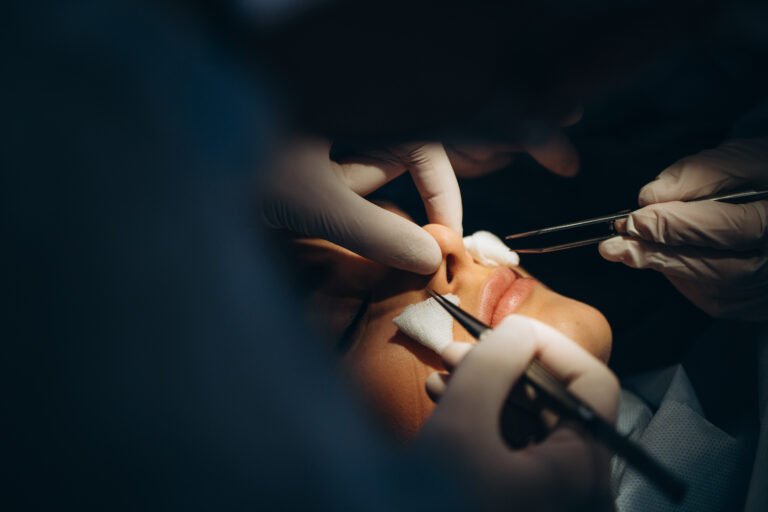
1. Surgical Treatment
Surgery is the primary treatment modality for JNA, particularly for tumors confined to the nasal cavity, sinuses, and skull base.
- Endoscopic Surgery:
- Minimally invasive techniques are now preferred for early-stage (I-II) tumors.
- Endoscopic approaches allow access to the tumor through the nostrils, reducing recovery time and avoiding facial incisions.
- Outcomes are favorable, with reduced blood loss compared to open surgery.
- Open Surgery:
- For advanced (Stage III-IV) tumors with significant skull base or intracranial involvement, open techniques such as craniofacial resection may be necessary.
- These surgeries are more invasive but offer better access for removing large or complex tumors.
2. Preoperative Embolization
- Embolization is often performed 24-48 hours before surgery to reduce blood loss by blocking the arteries supplying the tumor.
- It involves injecting particles through a catheter into the external carotid artery to prevent excessive intraoperative bleeding.
3. Radiotherapy
- Radiation therapy is reserved for tumors that are inoperable or recur after surgery. It may also be considered for advanced cases with intracranial involvement.
- Newer techniques, such as stereotactic radiosurgery (e.g., Gamma Knife), allow for targeted delivery of radiation to reduce the risk of damage to surrounding tissues.
4. Chemotherapy and Hormonal Therapy
- Chemotherapy is rarely used but may be employed for recurrent or unresectable tumors.
- Experimental treatments with anti-androgen therapy are being explored to target the hormonal factors driving the tumor’s growth.
Prognosis and Recurrence
- Recurrence rates vary between 10-40%, with higher rates seen in advanced-stage tumors.
- Most recurrences occur within the first two years after surgery, emphasizing the need for regular follow-up with imaging studies.
- Long-term prognosis is generally good for early-stage tumors treated surgically, but patients with advanced tumors involving the brain or orbit may experience complications.
Conclusion
Juvenile nasopharyngeal angiofibroma is a rare but challenging tumor that requires a combination of clinical expertise and advanced surgical techniques for optimal management. Advances in endoscopic surgery and preoperative embolization have improved patient outcomes by reducing complications and recovery times. However, the potential for recurrence necessitates careful follow-up, and new therapies continue to be explored to address the more advanced and recurrent cases.